Cornea
The cornea is a clear dome shaped structure at the front of the eye. Along with the lens of the eye, the cornea focuses light onto the retina to facilitate vision. Both the shape and the clarity of the cornea are essential to its function. Corneal diseases affect either the shape or clarity of this structure, resulting in challenges with vision.
Keratoconus
In keratoconus, the shape of the cornea is altered. Over time, the cornea becomes thin and steepens, or protrudes forward into the form of a cone. This change of shape causes light entering the eye to be poorly focused. As keratoconus progresses, vision declines, the cornea can become scarred, and patients may experience painful episodes of swelling known as corneal hydrops.
Management of keratoconus focuses on two objectives: preventing progression and optimizing vision. The major risk factor for and even cause of keratoconus is eye rubbing. Ceasing to rub and apply pressure to the eye is essential. In many cases, a procedure known as crosslinking is performed to stop progression. Rehabilitation of vision may require glasses or specialty contact lenses. In some cases, a device called Intacs is used to help flatten the cornea to improve vision in glasses or assist with the fit of contact lenses. In severe cases of keratoconus, a corneal transplant may be required.
Other conditions similar to keratoconus also require monitoring and care. These include pellucid marginal degeneration and ectasia, or weakening, after prior corneal surgery.
Corneal Collagen Crosslinking
Fuchs Corneal Dystrophy

Healthy corneal endothelial cells.
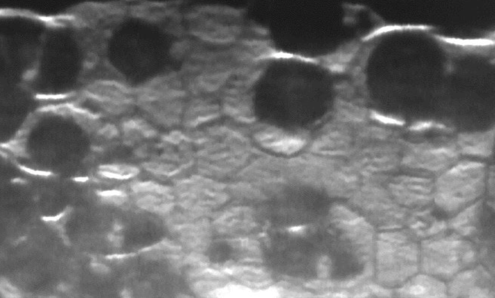
Fuchs dystrophy with loss of endothelial cells.
Corneal Transplantation
- Penetrating Keratoplasty (PKP)
- Descemet Membrane Endothelial Keratoplasty (DMEK)
- Descemet Stripping Endothelial Keratoplasty (DSEK)
- Deep Anterior Lamellar Keratoplasty (DALK)
This type of transplant is also known as a “full thickness” transplant. In this procedure all 5 layers of the cornea are removed. The donor cornea used for transplantation then also consists of all 5 layers, providing a complete or full thickness transplant.
Many corneal diseases only affect the innermost linings of the cornea. The most common of these conditions is Fuchs Corneal Dystrophy but there are others. Typically in these cases only the inner lining of cells needs to be replaced. A DMEK transplant replaces the least amount of corneal tissue relative to other transplantation techniques.
Other Corneal Conditions
Many other conditions can affect the cornea, thereby reducing vision. Infection, scarring, tumors, and inflammation may present in the cornea alone or secondary to other systemic medical conditions. At Glacier Eye Clinic we will do our best to assist you in diagnosing, treating, and coordinating with your other medical doctors to take care of your vision.
Additional information about the cornea and corneal disease may be found on the National Eye Institute website.